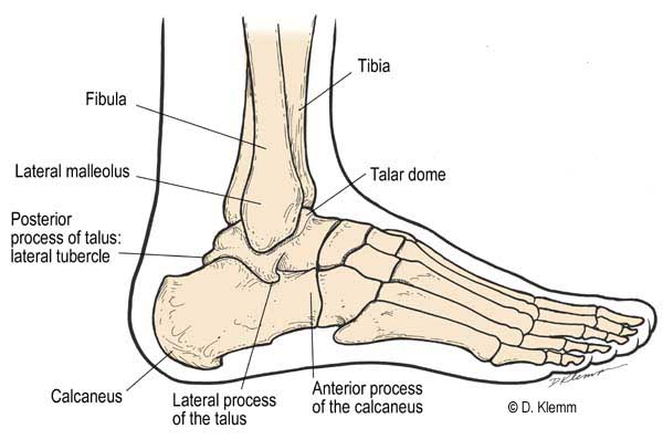
The Talus and Calcaneous are located on the posterior aspect of the foot. The talus consists of the body, neck and head and articulates with the calcaneous and navicular. It is only the foot bone that articulates with the fibula and tibia malleoli, and it consists of posterior, lateral and medial facets.
During walking, the talus initially bears the entire weight of the body as the person plants his or her foot. Approximately half the body's weight then is transmitted to the calcaneous, the largest and strongest foot bone. The calcaneous forms the heel and gives the support to the talus through three facets that articulate with the talus.
The foot bones are held together by ligaments and tendons that help form a two-way arch system consisting of longitudinal ( i.e. lengthwise) and lateral (i.e. transverse) arches. These arches enable the foot to support the weight of the human body and provide leverage during walking. The arches of the foot are not rigid they yield as weight is applied and spring back when weight is lifted. Strong fibrous structures normally hold the foot bones in their arched positions, but frequently these supporting structures weaken and result in foot deformities ( e.g. flatfoot).
The talocalcaneal joint has three articulations between the anterior, middle and posterior facets of the calcaneous and talus. These articulations function as single-unit, multi axial joint that allows the foot 40 deg of supination and pronation. Chopart's joint consists of articulations between the calcaneocuboid and talonavicular joints. The mid foot is unlocked when the hind foot is in valgus, therefore, surgeons slightly pronate the calcaneous when they fuse the tibiocalcaneal joint. |